This article was last reviewed on
This article waslast modified on 24 January 2020.
Overview of the heart

The heart is a muscular, fist-sized organ located within your chest in a space between the two lungs (mediastinum). The heart continuously pumps blood, beating as many as 100,000 times a day. The blood that the heart moves carries oxygen and nutrients throughout the body and transports carbon dioxide and other wastes to the lungs, kidneys, and liver for removal.

The heart is supplied with oxygen through a set of coronary arteries and veins. The heart is also an endocrine organ that produces the hormones atrial natriuretic peptide (ANP) and B-type natriuretic peptide (BNP), which coordinate heart function with blood vessels and the kidneys.

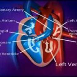
The heart is essentially hollow with two halves divided vertically by a septum, and each side of the heart has two internal chambers – an atrium on top and a ventricle on the bottom.

  • Blood returning from the body (oxygen-poor) through the veins enters the right side of the heart through the right atrium and is pumped by the right ventricle to the lungs, where carbon dioxide is released and oxygen is taken up.
  • This oxygenated blood from the lungs is returned to the left atrium and is pumped by the left ventricle into arteries that carry it throughout the body. Four heart valves regulate the direction and flow of blood through the chambers of the heart. It is their opening and shutting that gives the heart its typical "lub-dub" beat. The heart has an electrical system that controls the rate and rhythm of the heartbeat set by special heart cells called the sinoatrial node or the pacemaker.

The heart muscle itself is called the myocardium. The endocardium is a membrane that lines the chambers of the heart and the valves. The outside of the heart is encased by the pericardium – a layered membrane that is fibrous on the outside and serous (fluid-secreting) on the inside. The pericardium forms a protective barrier around the heart and allows it to beat in a virtually friction-free environment.


Accordion Title
About Heart Disease
  • What is heart disease?

    Heart disease is a general term that refers to a variety of acute and chronic medical conditions that affect one or more of the components of the heart. Diseases affecting the heart may affect the structure (structural) or how well it works (functional). Anything that damages the heart or decreases the heart’s supply of oxygen makes it less efficient, reduces its ability to fill and pump, and disrupts its coordinating relationship with blood vessels and the kidneys, so harming not only the heart but the rest of the body as well.

  • Signs and Symptoms

    Heart diseases may be acute or chronic and short-lived, relatively stable or progressive. They may cause a variety of signs and symptoms. Patients with early heart disease may experience few or vague symptoms, such as fatigue, shortness of breath with or without exercise, dizziness and/or nausea; however, these symptoms do not indicate any particular type of heart disease and may also be seen in a variety of other conditions. Other conditions associated with heart disease include:

    • Arrhythmia – an irregular heartbeat
    • Embolism – blockage of a blood vessel by material that has travelled from a distant body site, most often a blood clot, but it can be due to fat or air or even amniotic fluid and cells. A thrombus is a blood clot formed at that location, coronary artery thrombosis is another cause of coronary artery blockage
    • Inability to keep up with increased demands for oxygen and clearance of waste products, such as during physical activity
    • Infarction – death of muscle cells due to blockage of blood flow leading to scarring
    • Insufficient contraction – not emptying or filling completely
    • Pain, frequently due to ischaemia – a lack of sufficient oxygen because of reduced blood flow
    • Valve prolapse – part of the heart valve protrudes into the atrium, preventing a tight seal, which may lead to regurgitation and an increased risk of endocarditis
    • Valve regurgitation – a backflow of blood causing increased pressure in the blood vessels of the lungs and liver
    • Valve stenosis – narrowing of the opening which affects blood flow
    • Ventricular hypertrophy – increased thickness of the walls of the heart, causing a decrease in the size of the chambers and also a decrease in the flexibility of the heart

    Heart diseases may be due to:

  • Common Heart Diseases

    Coronary artery disease (CAD) causing coronary heart disease (CHD) is the most common form of heart disease. It is part of a general cardiovascular disease (CVD) – a narrowing of arteries in the heart and throughout the body over time due to a build-up of fatty inflammatory deposits that form plaques (atherosclerosis). This narrowing can significantly limit the amount of blood carried by the arteries and decrease the amount of oxygen supplied to the tissues. As the coronary blood vessels gradually narrow, symptoms do not appear until most of the blood flow is lost to an area of the heart, when it can cause intermittent chest pain upon exercise (angina) that worsens in frequency and severity over time potentially causing death of an area of the myocardium or heart attack (myocardial infarction). These forms of acute onset of chest pain are termed the acute coronary syndrome.

    Heart failure, usually called congestive heart failure (CHF), is when the heart has become less effective at circulating blood and less able to completely fill or empty the chambers. As a result blood backs up into the veins of the legs, lungs and liver, causing swelling, shortness of breath and fatigue. Any gradual damage to the heart can lead to heart failure over time; most commonly it is due to coronary artery disease, high blood pressure (hypertension) or heart valve damage (born with an abnormal valve or damaged later by an infection) or a previous heart attack. If the cause is temporary, heart failure may also be temporary; however it is usually a chronic and progressive condition.

    Cardiomyopathy is an abnormality of the heart muscle. One or more chambers of the heart may dilate (dilated cardiomyopathy), causing an enlargement in the size of one of the chambers of the heart. In other cases, one or more of walls of the heart may thicken (hypertrophic cardiomyopathy). Occasionally, cardiomyopathy may also be due to abnormal material accumulating in the wall of the heart, reducing the flexibility of the walls of the ventricles (restrictive cardiomyopathy). Cardiomyopathy may be due to decreased blood flow to the heart, exposure to chemicals that damage the heart (such as alcohol and cocaine, and medical drugs e.g. chemotherapy), to inherited muscle problems, to potassium or thiamine deficiency or it may have no obvious cause (called idiopathic cardiomyopathy).

    Myocarditis refers to inflammation of the muscle of the heart. It usually presents with an acute onset of shortness of breath or an irregular heartbeat, and can cause heart failure to develop quickly. It is often due to a viral infection.

    Pericardial disease is a disease of the sac surrounding the heart. It may be due to a bacterial or fungal infection, trauma, an autoimmune process, cancer or a previous heart attack. Infections of the pericardium may quickly become serious if left untreated. Pericarditis, an inflammation of the pericardium, may cause increased friction “rub” and pain in the chest.

    Congenital heart disease, which arises during fetal development, may affect any part of the heart. The amount of dysfunction depends on what form the abnormality takes and the extent to which it alters the shape, integrity and function of the heart.

    Endocarditis is an inflammation of the membrane lining the heart and heart valves. It may be due to a microorganism or to an autoimmune process. When it is due to an infection, it is often difficult to treat.

  • Tests

    The goals of testing for heart disease are to distinguish between symptoms that are heart-related and those that are due to another condition.  Testing is requested to help determine which heart disease is present, to determine whether the disorder is acute or chronic, to monitor a cardiac event that is in progress – such as a heart attack and to determine the severity and extent of the disease. 

    Patients presenting to the casualty department with an acute coronary syndrome – a group of symptoms that suggest heart injury due to lack of blood flow – are evaluated with laboratory blood tests and other tests. These are used to determine the cause of the pain and the severity of the condition. Since some treatments for a heart attack must be given within a short period of time to minimize heart damage, an accurate diagnosis must be quickly confirmed.

    Heart disease that is causing few symptoms may be detected during a visit to the doctor for nonspecific symptoms such as fatigue. Healthcare professionals may request a variety of blood and other tests to investigate possible causes for the patient’s symptoms.

    Testing patients for existing heart disease is not the same as cardiac risk testing. Cardiac risk testing is performed to screen asymptomatic people to help determine their risk of developing coronary heart disease.

    Laboratory Blood Tests
    Cardiac biomarkers are proteins that are released when muscle cells are damaged. Tests include:

    • Troponin - used to detect ischaemic damage (lack of blood flow) to the heart in suspected acute coronary syndrome
    • BNP – used to detect and monitor heart failure (stretch)

    More general blood tests that may be requested:

    • Blood Gases – performed to evaluate oxygen, carbon dioxide, and pH levels
    • Electrolytes – tests that evaluate the body’s electrolyte balance and kidney function
    • Full Blood Count (FBC) – looks at blood cells, checks for anaemia (low haemoglobin can cause ischaemia even though coronary blood vessels are clear and open)

    Other Evaluations
    A range of other evaluations and tests are used to assess chest pain and other symptoms. These include:

    • A medical history, including an evaluation of risk factors such as age, CAD, diabetes, and smoking
    • A physical examination
    • An electrocardiogram (ECG) – a test that looks at the heart’s electrical activity and rhythm
    • Echocardiography – ultrasound imaging of the heart

    Based on the findings of these tests, other procedures may be necessary, including:

    • Stress testing
    • Chest X-ray
    • Computerised tomography (CT) scan including coronary calcium scan or coronary angiography
    • Continuous ECG monitoring (sometimes also called Holter monitoring or 24 hour tape) – the patient wears a monitor that evaluates heart rhythm over a period of time
    • Magnetic resonance imaging (MRI)
    • Positron emission tomography (PET)
    • Radionuclide imaging
    • Cardiac catheterisation – in this procedure, a thin flexible tube is inserted into an artery in the leg or arm and threaded up to the coronary arteries to evaluate blood flow and pressure in the heart and the status of the arteries in the heart
    • Coronary angiography – X-rays of arteries using a radiopaque dye to help diagnose CAD; this procedure is performed during coronary catheterisation or CT scan


  • Treatments

    Treatments for heart disease depend on the condition and its severity. Acute conditions, such as a heart attack, require prompt medical intervention to minimize heart damage. For chronic conditions, doctors may recommend that affected patients modify their diets, lose excess weight, exercise (under supervision), and quit smoking. Conditions such as hypertension and diabetes should be controlled to minimise their effect on the heart. Heart failure often requires treatment with low salt diet and diuretics (to try to reduce the amount of fluid accumulation) as well as drugs such as digoxin to improve the function of the heart muscle.

    Drugs may be necessary to help manage symptoms and control heart diseases. Surgical procedures may be required to bypass blocked arteries, replace defective heart valves or correct congenital abnormalities. Those with infections may need to take antimicrobial drugs. Electrical devices to control or reset heart rhythm and help the heart contract can be inserted e.g. pacemakers. In very rare cases the organ may need to be replaced e.g. heart transplant or combined heart and lung transplant.

    New drugs, procedures, and guidelines for addressing heart disease continue to be developed. Patients should talk to their doctor about the best treatment options for their condition.