This article was last reviewed on
This article waslast modified on 7 March 2022.
What are they?

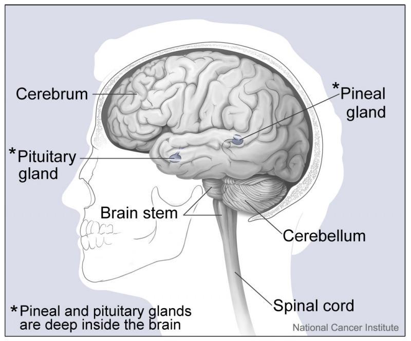
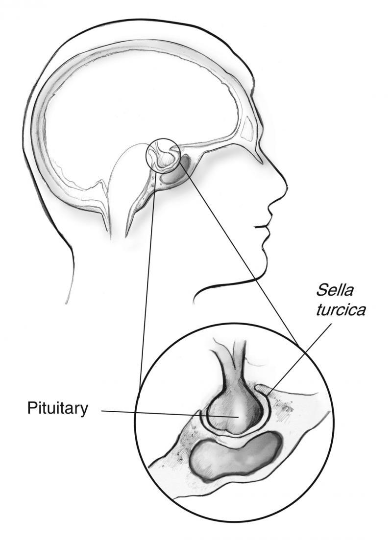
The pituitary is a pea-sized gland that is attached to an area at the base of the brain called the hypothalamus. The pituitary gland is part of the endocrine system; a system of glands that produce and regulate hormones. The hypothalamus communicates with the rest of the brain and nervous system. It senses the body’s need for more or less of a particular hormone and controls the amount of hormone released from the pituitary. Pituitary disorders are characterised by an excess or a deficiency in one or more of the hormones produced by the pituitary gland. When the disorder is caused by a pituitary tumour, symptoms such as headache and loss of vision may be present due to compression of the surrounding tissues.

The pituitary consists of a front (anterior) and back (posterior) portion. In the anterior pituitary, growth hormone (GH), adrenocorticotrophic hormone (ACTH), thyroid-stimulating hormone (TSH), luteinising hormone (LH), follicle-stimulating hormone (FSH) and prolactin are produced. They affect particular “target” tissues throughout the body, including the adrenal glands, thyroid gland, ovaries (women), and testes (men). Oxytocin and antidiuretic hormone (ADH) are produced in the hypothalamus and stored in the posterior pituitary for release. Some of the normal functions of these hormones are described below.

  • GH: Regulates bone growth and muscle mass.
  • ACTH: Regulates the body’s response to stress by stimulating cortisol production by the adrenal glands.
  • TSH: Stimulates the thyroid gland to produce thyroid hormones, which regulate the rate at which the body uses energy (the metabolic rate).
  • LH and FSH: Required for the development of sexual characteristics at puberty and fertility.
  • Prolactin: Stimulates breast milk production after childbirth.
  • ADH: Controls the amount of water that the kidneys excrete, which in turn helps regulate the balance of water in the body.
  • Oxytocin: Stimulates the contraction of the uterus during and after childbirth and is responsible for stimulating the release of milk during breastfeeding.
Accordion Title
About Pituitary Disorders
  • Causes

    What can go wrong?
    The growth of a tumour is the most common problem with the pituitary. While most are benign (pituitary adenoma), they can produce excessive amounts of a specific pituitary hormone, reduce the production of other pituitary hormones, and compress surrounding tissues. Blood vessels and the optic nerves are in close proximity to the pituitary gland. Pressure from a tumour can cause vision problems (including loss of vision), headaches, fatigue, weakness, and seizures, as well as a host of signs and symptoms related to decreased or increased hormone production. The hormone deficiencies and excesses from these disorders can produce a variety of symptoms depending on which hormones and target tissues are affected. Symptoms may not be present and tumours can be found when imaging is performed for other reasons (incidentalomas).

    Other causes of pituitary disorders include:

    • Surgical or radiation treatment impacting the pituitary
    • Physical accident (trauma)
    • An impaired blood supply to the pituitary
    • Infection
    • Infiltration
    • An autoimmune disorder
    • Inherited genetic mutations
    • Congenital disorders (present from birth)
    • Malignant tumours (rare)
    • Causes that are not yet well understood

    Pituitary hormone production can be affected when the hypothalamus is not functioning properly. Excess or deficient hormone production by the pituitary may also occur if the glands “downstream” from it are not functioning properly. For example, normally the hypothalamus detects low levels of thyroid hormone in the blood and stimulates the pituitary to produce TSH. TSH in turn stimulates the thyroid gland to produce thyroid hormones. If the thyroid gland cannot produce enough thyroid hormones, then blood thyroid hormone levels will remain below normal even though the hypothalamus and pituitary are promoting production by increasing the amount of TSH in the blood. This results in very high levels of TSH and abnormally low levels of thyroid hormones.

  • Examples of Disorders
    • Pituitary Tumours: May be hormone-secreting or non-secreting. Most are benign, but rarely they are malignant. May cause visual disturbances and headaches as they grow and compress surrounding tissues. Often results in excessive amounts of one pituitary hormone and decreases in others.
    • Growth Hormone Deficiency: From a variety of causes. In children it causes delayed growth and short stature. In adults it can lead to muscular weakness, fatigue, decreased bone mass, and obesity.
    • LH and FSH (gonadotrophin deficiency): Causes low testosterone, decreased libido, and erectile dysfunction in males and menstrual disorders in females.
    • Hyperprolactinaemia: A pituitary tumour that secretes prolactin or a tumour that prevents the regulation of prolactin production. In women can cause galactorrhoea (milk secretion from the breasts) and amenorrhoea (loss of menstrual cycles), and in men decreased sex drive and impotence. Some tumours secrete growth hormone as well as prolactin.
    • Hypopituitarism: From a variety of causes including tumours (benign and malignant), head trauma, decreased pituitary blood supply, stroke / haemorrhage, infection (e.g. tuberculosis), infiltration (e.g. sarcoidosis), an autoimmune disorder, radiation therapy, surgical removal of the pituitary, a side effect of pituitary surgery, hemochromatosis (iron overload), congenital or genetic diseases. Results in a general decrease in pituitary hormone production. As well as the hormone deficiencies already mentioned, low cortisol (ACTH) and hypothyroidism (low TSH) can occur. Symptoms may relate to the hormone deficiency, tissue compression by the tumour or be non-specific e.g. fatigue.
    • Acromegaly and gigantism: Excess growth hormone production, usually due to a tumour. When it occurs in childhood, it causes gigantism associated with excessive bone growth and abnormally tall stature. In adults, it causes acromegaly, with increases in bone thickness, coarsened facial features, enlarged hands and feet, carpal tunnel syndrome (aching, numbness and tingling in hand), headaches, loss of vision, sweating, sleep apnoea (breathing problems at night associated with snoring), fatigue, and cardiovascular disease e.g. hypertension. Hyperprolactinaemia may also be present. Menstrual disturbance may occur in women and sexual dysfunction in men.
    • Cushing Disease: Caused by a pituitary tumour that produces excess ACTH and leads to excess exposure by the body to the adrenal gland hormone cortisol. Symptoms vary but include: upper body obesity, a rounded face, thin skin, pink streaks on the abdomen, muscular weakness, osteoporosis, high blood sugar, and high blood pressure.
    • Diabetes Insipidus: Decreased production of ADH by the hypothalamus. The patient’s kidneys don’t conserve water and concentrate urine; patient is thirsty and has frequent, dilute urination.
    • Craniopharyngioma: A type of brain tumour that develops close to the pituitary gland; roughly half occur in children and adolescents with the rest occurring in adults over 20 years old. Benign but may put pressure on the pituitary, causing delayed growth in children, visual disturbances / headaches in adults and hypopituitarism.
    • Multiple Endocrine Neoplasia Type 1 (MEN1): An inherited genetic mutation that increases the risk of developing tumours in the pituitary and in other endocrine glands (parathyroid and pancreas).
    • Pituitary Infarction: Restricted blood supply to the pituitary gland. May cause gland tissue damage and lead to hypopituitarism.
    • Sheehan Syndrome: This is a very rare condition caused by pituitary infarction following severe blood loss during childbirth. Results in hypopituitarism.
    • Empty Sella Syndrome: The sella is the structure that surrounds the pituitary gland. It may increase in size and put pressure on the pituitary. Rarely, the pituitary gland shrinks in response and hormone production decreases, leading to hypopituitarism.
    • Kallman Syndrome: Deficient release of GnRH (gonadotropin-releasing hormone) from the hypothalamus leads to lack of FSH and LH production. Causes delayed or absent puberty, can be associated with no sense of smell, and is more common in males.
    • Nelson Syndrome: May result when both adrenal glands are removed as part of the treatment for Cushing Disease. A pituitary tumour develops that produces ACTH and can cause darkening of the skin due to increased production of melanocyte stimulating hormone (MSH).
    • TSH-secreting pituitary adenomas (rare): Can cause hyperthyroidism through increased TSH secretion. Signs / symptoms include goitre, palpitations, tremor, heat intolerance, visual disturbances, headaches and menstrual disturbances (women).
    • Mixed cell adenomas: Excess secretion of multiple hormones e.g. prolactin and growth hormone. Can involve any combination of cells/hormones.
  • Tests

    The goals of pituitary disorder testing are to detect excess or deficient hormone production, determine the cause, and evaluate the severity of the condition. Testing frequently includes measurements of both the hormones that the pituitary produces (e.g. TSH), and the hormones of other endocrine glands that the pituitary is responsible for stimulating (e.g. the thyroid hormone thyroxine).

    Pituitary hormones are released as needed. Concentrations of some hormones (such as TSH) are relatively constant in the blood, whilst others vary over the course of a day, (GH), or over a cycle (such as FSH and LH during the menstrual cycle). The concentration of a hormone may also be dependent on specific situations. For example, prolactin is raised in women that are breast feeding. Sometimes, because of these variations, the result from a single blood test does not give enough information. Tests which involve the patient taking medication to either suppress or stimulate hormone production can be used to detect hormonal excess or deficiency, respectively. Related tests may also be used as a marker of the hormone under investigation. For example, measurement of IGF-1 (insulin-like growth factor-1) reflects total growth hormone production.

    Laboratory Tests

    • Prolactin
    • LH and FSH (with oestradiol in females and 9am testosterone in males)
    • TSH and Free, T4 (Thyroxine)
    • ACTH, 9am Cortisol (if deficiency suspected), 24h urine cortisol / overnight dexamethasone suppression / midnight salivary cortisol (if cortisol excess is suspected)
    • IGF-1 (±GH)
    • Urine and serum osmolality (for investigation of Diabetes Insipidus)

    Non-Laboratory Tests

    • MRI (magnetic resonance imaging)
    • CT (computed tomography) scan
    • Visual field testing

    Once a diagnosis has been made, testing of pituitary function may be requested at regular intervals to monitor the effectiveness of the treatment given. Such tests are necessary as treatments may not completely resolve the condition. Furthermore, treatment may cause additional pituitary dysfunction either immediately (following surgery) or in the future (following radiation). Long term monitoring may also be necessary if a person has an inherited condition that presents a lifetime risk of developing a pituitary disorder.

  • Treatments

    Treatments vary depending on the reason for the pituitary disorder.

    When a pituitary tumour is present, surgery may be performed to remove the tumour. The pituitary gland is accessed through the nose in a procedure called transsphenoidal surgery. This procedure is delicate and requires a surgeon that is experienced in the procedure.

    Medicines may be used to shrink pituitary tumours to reduce compression symptoms, and sometimes to make pituitary surgery easier to perform. Medication may also be used to block excessive hormone production. Hormone replacement may be used to supplement deficient hormones.

    Targeted radiation therapy may be used in conjunction with other treatments to decrease tumour size and excess hormone production (after surgery and medical treatments have been exhausted).

    Patients should work with their doctor and/or an endocrinologist (a doctor that specialises in treating disorders of the endocrine system), to determine the best course of treatment(s) for them.